The eukaryotic cell cycle coordinates growth, DNA replication, chromosome segregation and cytokinesis so that daughter cells inherit complete genomes.
Key takeaways
- The cell cycle is commonly divided into G1, S, G2 and M phases, with DNA replication restricted to S phase.
- Mitosis segregates duplicated chromosomes; cytokinesis divides the cytoplasm.
- Checkpoint systems delay progression when DNA is damaged or chromosome attachment is incomplete.
In this article
- The cell cycle is more than mitosis
- DNA must be replicated first
- Chromosome cohesion and condensation make segregation possible
- Building the mitotic spindle and checking attachment
- From metaphase to anaphase
- Cytokinesis
- Cyclins and CDKs drive the transitions
- Checkpoints monitor completion, not perfection
- Cell-cycle failure is central to cancer biology
- Not every cell is continuously cycling
- Mitosis varies in detail across eukaryotes
The cell cycle is more than mitosis
Mitosis is the visually dramatic part of cell division, but most proliferating cells spend the majority of their cycle in interphase. During G1 the cell grows and responds to environmental signals. DNA is duplicated during S phase. G2 provides additional growth and preparation before M phase.
Some cells can leave the active cycle and enter a quiescent state often called G0. In multicellular organisms, whether a cell divides depends on its tissue, developmental state and extracellular signals.
DNA must be replicated first
Before chromosomes can be segregated, each DNA molecule is copied. The two copies remain associated as sister chromatids. Proteins organize and compact the chromosomes so that long DNA molecules can be moved without becoming hopelessly entangled.
Replication errors and DNA damage can activate signaling pathways that delay later stages. These controls reduce the chance that damaged or incomplete genomes will be passed to daughter cells.
Chromosome cohesion and condensation make segregation possible
Duplicated DNA molecules would be difficult to segregate if they remained as long, loosely organized chromatin fibers. Cohesin protein complexes help hold sister chromatids together after replication, while condensin complexes and other chromosome-associated proteins promote the compact architecture of mitotic chromosomes. At anaphase, regulated cleavage of cohesin allows sister chromatids to separate only after correct spindle attachment has been established.[4]
Building the mitotic spindle and checking attachment
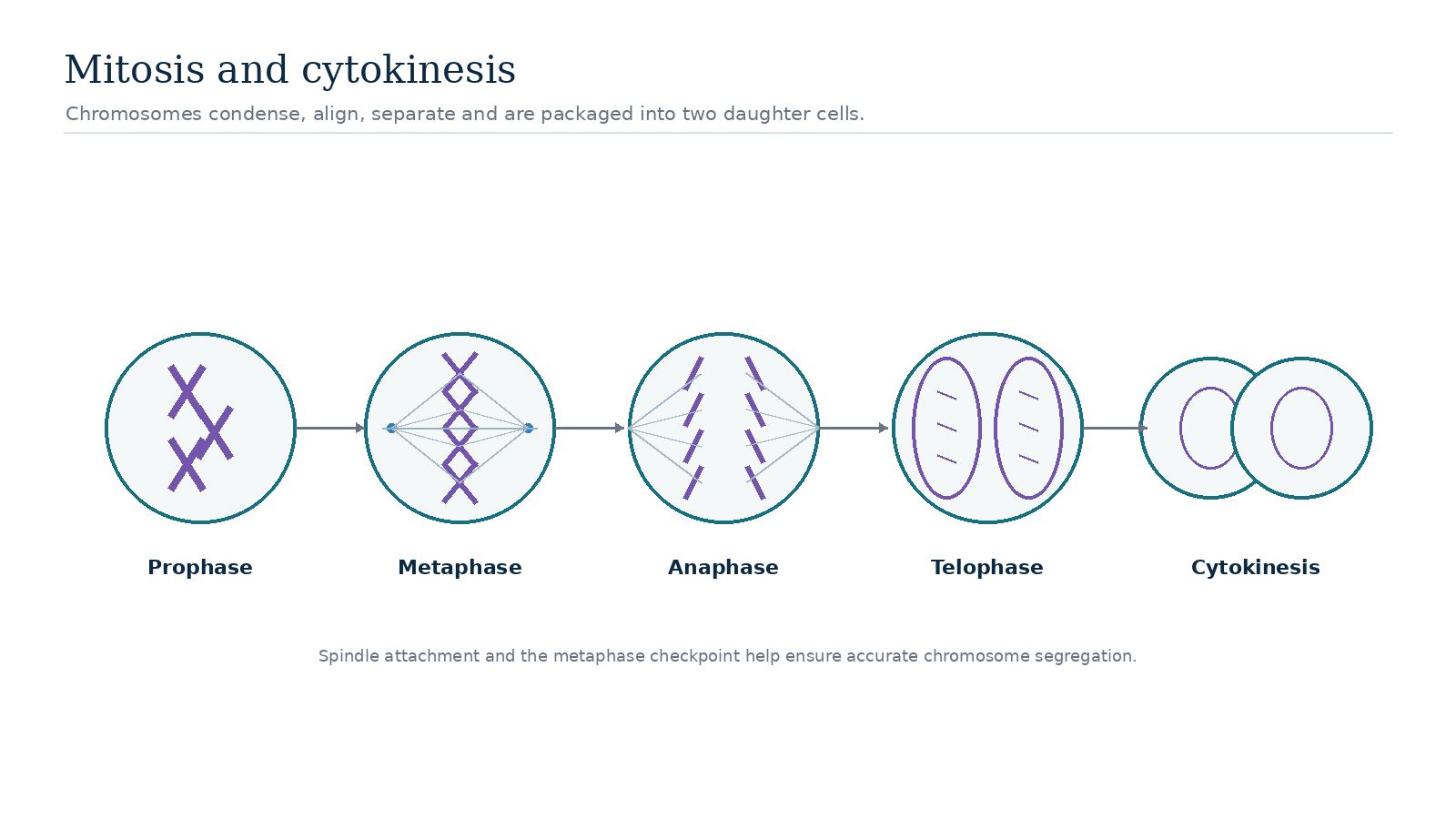
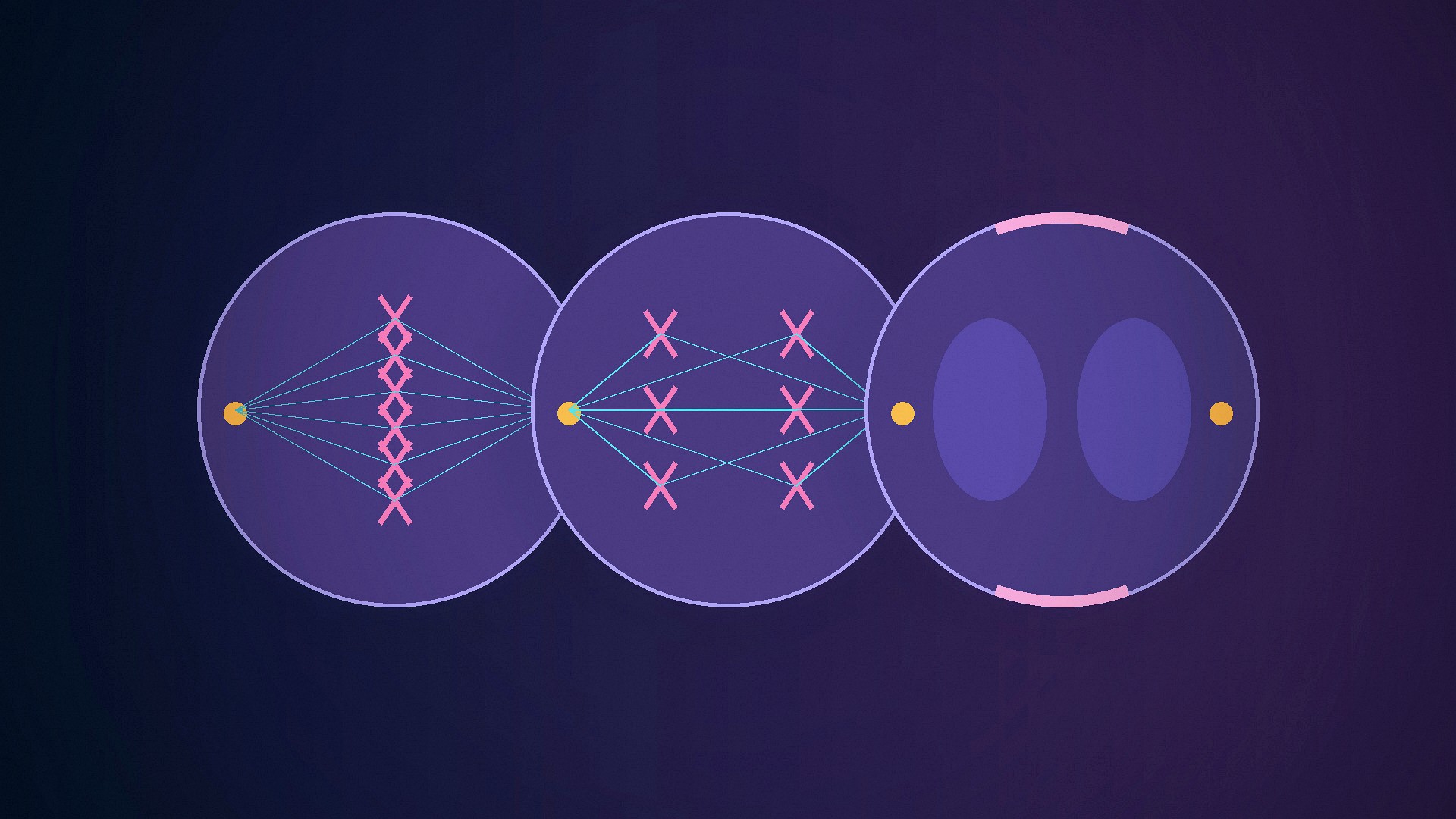
During mitosis, microtubules assemble into a spindle that connects duplicated chromosomes with opposite spindle poles. Protein structures called kinetochores form at centromeric regions and provide attachment sites for spindle microtubules. Correct bipolar attachment creates tension and positions chromosomes near the cell’s equator during metaphase.[4]
The spindle-assembly checkpoint delays anaphase while chromosomes remain unattached or incorrectly attached. It reduces the probability of chromosome-segregation errors but cannot guarantee a perfect division every time. Persistent segregation errors can produce aneuploid daughter cells with abnormal chromosome numbers.[2]
From metaphase to anaphase
When the cell satisfies the checkpoint, cohesion between sister chromatids is released. The chromatids move toward opposite poles during anaphase while the spindle reorganizes and elongates.
By telophase, chromosomes arrive at the poles, begin to decondense and are enclosed by re-forming nuclear envelopes. This completes nuclear division.
Cytokinesis
Animal cells usually divide their cytoplasm using a contractile ring containing actin and myosin. The ring constricts the cell membrane between the new nuclei until the cells separate. Plant cells cannot pinch inward through a rigid cell wall, so they build a cell plate that develops into new separating walls and membranes.
Mitosis and cytokinesis are coordinated but distinct processes. A failure in chromosome segregation can produce cells with abnormal chromosome numbers, while a failure in cytokinesis can create multinucleate cells.
Cyclins and CDKs drive the transitions
The cell cycle is controlled by cyclin-dependent kinases (CDKs), enzymes whose activity changes when they bind regulatory proteins called cyclins. Different cyclin–CDK combinations become active at different stages, phosphorylating proteins that commit the cell to DNA replication, prepare it for mitosis or trigger chromosome separation. Cyclin concentrations rise and fall because cells synthesize and destroy them in a regulated sequence.[1]
This control system makes the cycle directional. Once key proteins are degraded at the metaphase-to-anaphase transition, the cell cannot simply reverse chromosome segregation and return to metaphase. Similar irreversible steps help ensure that DNA is replicated once per cycle and that mitosis follows replication rather than preceding it.
Checkpoints monitor completion, not perfection
Checkpoints delay progression when essential conditions are not met. DNA-damage signaling can slow the G1/S or G2/M transition, giving repair systems more time to work. The spindle-assembly checkpoint prevents anaphase while chromosomes remain improperly attached to spindle microtubules. These systems reduce errors, but they cannot guarantee a perfect division every time.[2]
Cell-cycle failure is central to cancer biology
Genes controlling proliferation, checkpoints and programmed cell death are frequent targets of cancer-associated mutations. A cell that divides despite DNA damage can accumulate additional alterations; a cell that ignores anti-growth signals can expand when it should remain quiescent. Cancer is not simply “fast mitosis,” but loss of multiple layers of normal control over growth, genome maintenance and tissue organization.[3]
For the same reason, many anticancer drugs target processes required for cell division, including DNA synthesis or microtubule dynamics. These treatments can also affect healthy tissues with rapidly dividing cells, which helps explain some of their side effects.
Not every cell is continuously cycling
Many differentiated cells spend long periods outside the active cycle in a state often called G0. Some can re-enter the cycle after receiving appropriate signals; others are effectively post-mitotic under normal conditions. Stem and progenitor cells balance self-renewal with production of differentiated descendants, so their cell-cycle control is closely linked to tissue maintenance. This variation matters because the familiar G1–S–G2–M diagram is a framework for proliferating cells, not a claim that every cell in the body is always progressing toward its next division.
Cell-cycle length also varies enormously. Early embryonic cells can divide rapidly, while many adult cells divide rarely. The same molecular framework is tuned differently according to tissue type, developmental stage and external growth signals. Nutrient availability, cell size and contact with neighboring cells can also influence whether a cell commits to another division.
Mitosis varies in detail across eukaryotes
The familiar animal-cell diagram is not the only form of mitosis. Fungi and some other eukaryotes can retain much of the nuclear envelope during chromosome segregation, and organisms differ in how spindle poles are organized and how cytokinesis proceeds. Plant cells, for example, build a cell plate rather than pinching inward through a rigid cell wall.[5]
The conserved problem is the same: duplicate the genome once, establish attachments that separate sister chromatids to opposite sides and coordinate chromosome segregation with physical division of the cell. Evolution has modified the mechanics while preserving that core logic.[5]