DNA replication copies a cell’s genetic information before division. Here is how helicase, primase, DNA polymerase, ligase and proofreading systems coordinate the process.
Key takeaways
- DNA replication is semiconservative: each daughter DNA molecule contains one parental strand and one newly synthesized strand.
- DNA polymerases can extend DNA only in the 5′→3′ direction, which creates a continuously copied leading strand and a discontinuously copied lagging strand.
- Proofreading, repair and careful coordination at the replication fork keep the error rate extremely low.
In this article
- Why cells must copy DNA before division
- The replication fork: where copying happens
- Primers and DNA polymerase
- How the lagging strand is finished
- Why replication is so accurate
- What replication errors can mean
- Replication in chromosomes: origins, timing and chromosome ends
- Replication is coordinated with chromatin and DNA repair
- How scientists measure replication
Why cells must copy DNA before division
Every time a cell divides, each daughter cell needs a complete set of genetic instructions. In organisms whose cells contain chromosomes, that means the DNA must be duplicated before the chromosomes can be separated. Replication is therefore not simply a copying reaction; it is a coordinated sequence of molecular events that must preserve the order of billions of DNA building blocks while the chromosome is being opened, stabilized, copied and reassembled.
The underlying logic is simple. The two strands of a DNA double helix are complementary: adenine pairs with thymine, and guanine pairs with cytosine. When the strands separate, each can serve as a template for a new partner strand. The result is called semiconservative replication because each completed DNA double helix contains one strand from the original molecule and one newly synthesized strand.[4]
The replication fork: where copying happens
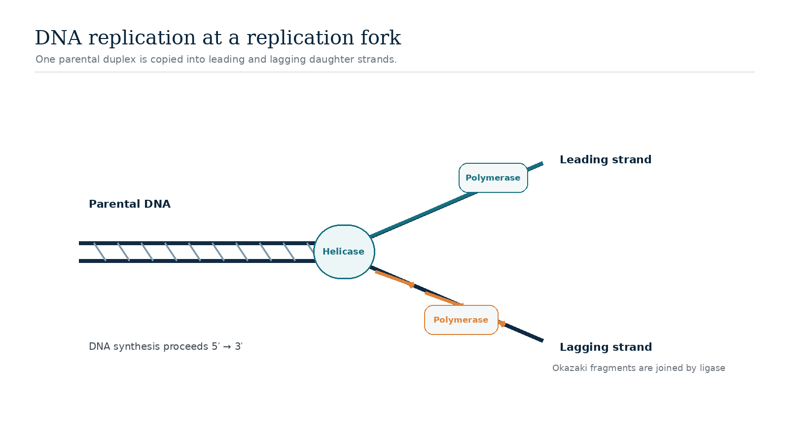
Replication begins at defined regions called origins. Proteins assemble there, locally unwind the DNA and create structures called replication forks. A helicase moves along DNA and separates the two parental strands. Single-strand binding proteins help prevent the exposed strands from snapping back together, while topoisomerases relieve twisting stress that builds ahead of the fork as the helix is opened.
The fork is not a single enzyme working in isolation. It is better understood as a moving molecular machine in which several proteins interact. Their timing matters: DNA must be opened quickly enough for polymerases to copy it, but exposed single-stranded DNA must also be protected because it is chemically vulnerable.
Primers and DNA polymerase
DNA polymerases catalyze the addition of DNA nucleotides to a growing strand, but they cannot normally start a new strand from nothing. A primase first builds a short RNA primer that provides a starting 3′ end. DNA polymerase can then extend from that primer by adding nucleotides that are complementary to the template.
Polymerases synthesize DNA only in the 5′→3′ direction. Because the two parental strands run in opposite orientations, the geometry of the fork creates two different copying strategies. On the leading strand, synthesis can proceed continuously in the same overall direction as fork movement. On the lagging strand, synthesis occurs in short stretches called Okazaki fragments. Each fragment begins with a new primer and is later joined to its neighbors.
How the lagging strand is finished
After an Okazaki fragment has been made, the RNA primer has to be removed and the resulting gap filled with DNA. Enzymes then process the junctions so that the sugar-phosphate backbone becomes continuous. DNA ligase performs the final sealing step by forming a phosphodiester bond between adjacent DNA fragments.
This repeated cycle of priming, extension, primer removal and ligation makes lagging-strand synthesis look more complicated than leading-strand synthesis, but both sides of the fork are tightly coordinated. In many organisms, multiple polymerase molecules and accessory proteins work together so that both templates are copied at roughly the same pace.
Why replication is so accurate
Base pairing provides the first level of accuracy, but polymerases also discriminate strongly between correct and incorrect nucleotides. Many replicative DNA polymerases have a proofreading activity: when the wrong nucleotide is inserted, the enzyme can move the newly made strand into a separate catalytic site, remove the mismatch and continue synthesis.
Additional mismatch-repair pathways can detect some errors that escape polymerase proofreading. No system is perfect, and occasional changes become mutations. Yet the combined effect of nucleotide selection, proofreading and repair reduces the frequency of permanent copying errors to a tiny fraction of the number of bases replicated.
What replication errors can mean
Most replication errors are corrected. Some that remain have little effect, while others can alter a gene or a regulatory region. In germ cells, a mutation can potentially be passed to future generations. In body cells, accumulated mutations can contribute to aging, clonal variation or diseases such as cancer if they affect genes that control growth, DNA repair or cell survival.[5]
Replication is therefore both a mechanism of biological continuity and a source of evolutionary variation. Its fidelity preserves genomes from one cell generation to the next, while the rare changes that survive repair provide some of the heritable variation on which evolution can act.
Replication in chromosomes: origins, timing and chromosome ends
Large chromosomes cannot be copied efficiently from a single starting point. Eukaryotic chromosomes therefore use many replication origins that activate in a regulated program during S phase of the cell cycle. Replication forks move outward from active origins until they meet forks arriving from neighboring regions. Licensing and checkpoint systems help ensure that each region is copied once and that cells do not enter chromosome segregation with extensive unfinished DNA.
Linear chromosomes create an additional problem at their ends because conventional lagging-strand synthesis cannot fully copy the extreme end of a template. Telomeres provide repetitive DNA that protects chromosome termini, and the enzyme telomerase can extend these repeats in cell types where it is active. These mechanisms connect replication biology to aging, cancer research and genome engineering, including technologies such as CRISPR.
Replication is coordinated with chromatin and DNA repair
In eukaryotic cells, copying DNA also means copying the chromosome’s packaging. DNA is wrapped around histone proteins in chromatin, so nucleosomes must be temporarily disrupted ahead of a replication fork and rebuilt behind it. Cells recycle many parental histones and add newly synthesized histones, helping daughter chromosomes regain an organized chromatin structure after the fork passes.[1]
Replication also intersects with DNA-repair systems. A polymerase can stall at damaged bases, tightly folded DNA or difficult repetitive sequences. Cells can stabilize a stalled fork, repair the obstacle or use specialized polymerases that tolerate certain lesions. These responses are important because a broken or collapsed fork can produce chromosome rearrangements rather than a simple one-letter mutation. The broader term replication stress describes situations in which DNA synthesis is slowed or disrupted, and persistent replication stress is common in many cancers.[2]
How scientists measure replication
Researchers study replication with biochemical assays, microscopy and genome-wide sequencing. Short pulses of labeled nucleotides can reveal where new DNA is being made, while sequencing-based methods can map active origins and the direction in which forks move. Structural techniques such as cryo-electron microscopy have also shown how helicases, polymerases and accessory proteins assemble into large complexes rather than acting as isolated enzymes.
These experiments reinforce an important point: the textbook replication fork is a useful simplification. In a living chromosome, synthesis is coupled to chromatin assembly, DNA repair, checkpoint signaling and the three-dimensional organization of the nucleus. The core chemistry is conserved, but the exact proteins and control systems differ between bacteria, archaea and eukaryotes.[3]