The brain integrates sensation, movement, memory, emotion and internal regulation through specialized but deeply interconnected neural systems.
Key takeaways
- The brain is organized into specialized regions, but most complex behaviors depend on networks that span multiple regions.
- Neurons communicate electrically within cells and chemically across synapses, while glial cells support, regulate and protect neural function.
- The cortex, basal ganglia, cerebellum, brainstem and limbic structures make distinct contributions that are coordinated continuously.
In this article
- A network, not a collection of isolated modules
- Neurons and synapses
- The cerebral cortex
- Deep brain structures
- Cerebellum and brainstem
- How the brain changes
- Glial cells support neural function
- Plasticity has limits as well as possibilities
- How scientists study brain activity and connectivity
- Blood flow is part of brain function
- Functional lateralization is real but often oversimplified
A network, not a collection of isolated modules

The human brain weighs only a small fraction of body mass, yet it coordinates sensation, movement, thought, memory, emotion and many automatic functions needed for survival. Textbook diagrams divide the brain into regions because anatomy matters, but no complex mental function belongs to a single isolated spot.
Instead, neural processing is distributed across networks. A task such as speaking requires sensory processing, memory, motor planning, movement control and feedback. Damage to one node can disrupt the network in characteristic ways, which is one reason neurological examination can reveal where an injury has occurred.
Neurons and synapses
Neurons are specialized cells that receive, integrate and transmit information. Electrical changes in the cell membrane can trigger an action potential that travels along an axon. At many synapses, the arriving signal causes neurotransmitters to be released and bind receptors on another cell.[1]
Synapses can strengthen or weaken over time. This plasticity is one of the cellular foundations of learning and adaptation. The brain also contains abundant glial cells, which support neurons, regulate the chemical environment, form myelin, participate in immune defense and influence synaptic function.
The cerebral cortex
The cerebral cortex is the folded outer layer of the cerebrum. Different cortical areas are associated with sensory processing, voluntary movement, language and higher cognitive functions. The frontal lobes contribute to planning, decision-making and motor control; parietal regions integrate body and spatial information; temporal regions are important for hearing and memory; and occipital regions are central to vision.[2]
These labels are useful but simplified. Information moves through reciprocal connections, and many functions recruit widely distributed circuits rather than following a one-way chain.
Deep brain structures
Below the cortex are structures that help regulate movement, motivation, memory and emotion. The basal ganglia participate in action selection and habit learning. The thalamus relays and coordinates much of the information traveling to the cortex. The hippocampal formation is important for forming certain kinds of new memories.
The hypothalamus links the nervous system with endocrine and autonomic regulation. It helps control temperature, hunger, thirst, circadian rhythms and hormonal signaling through interactions with the pituitary gland.
Cerebellum and brainstem
The cerebellum contains a very large share of the brain’s neurons and is essential for timing, coordination and motor learning. It continually compares intended movement with sensory feedback and helps refine ongoing actions. Research also links cerebellar circuits to non-motor functions.
The brainstem connects the brain with the spinal cord and contains networks involved in breathing, cardiovascular control, sleep-wake regulation and many cranial-nerve functions. Injury to the brainstem can therefore affect fundamental life-sustaining processes.
How the brain changes
The brain is dynamic across the lifespan. Development changes the number and strength of synaptic connections, experience reshapes circuits, and learning alters neural activity patterns. After injury, some functions can sometimes be reorganized through rehabilitation and plasticity, although the extent of recovery varies.
Brain disorders can arise from vascular disease, degeneration, abnormal electrical activity, infection, trauma, developmental differences, tumors and many other causes. General descriptions of brain anatomy cannot diagnose symptoms; persistent or sudden neurological symptoms require appropriate medical evaluation.
Glial cells support neural function
Neurons receive much of the attention, but glial cells are essential to brain function. Astrocytes help regulate the chemical environment around synapses and participate in signaling between neurons and blood vessels. Oligodendrocytes form myelin around many axons in the central nervous system, changing how rapidly signals travel. Microglia survey the tissue and respond to injury, infection and cellular debris. These cell types interact continuously with neurons, so healthy neural circuits depend on more than neurons alone.
Glial biology also changes across development, aging and disease. Loss of myelin can disrupt communication between regions, excessive or poorly regulated inflammation can injure tissue, and altered support of synapses can affect how circuits function. These examples are one reason modern neuroscience treats glia as active participants in nervous-system physiology rather than as structural filler between neurons.[3]
Plasticity has limits as well as possibilities
Synapses and networks change with experience, development and injury. This neuroplasticity supports learning and can contribute to recovery after damage, but it does not mean the brain can reorganize without constraint. Age, location and extent of injury, prior learning and rehabilitation all influence how much function can be recovered. Plasticity is a biological property of neural circuits, not evidence that every lost function can simply be reassigned elsewhere.[3]
How scientists study brain activity and connectivity
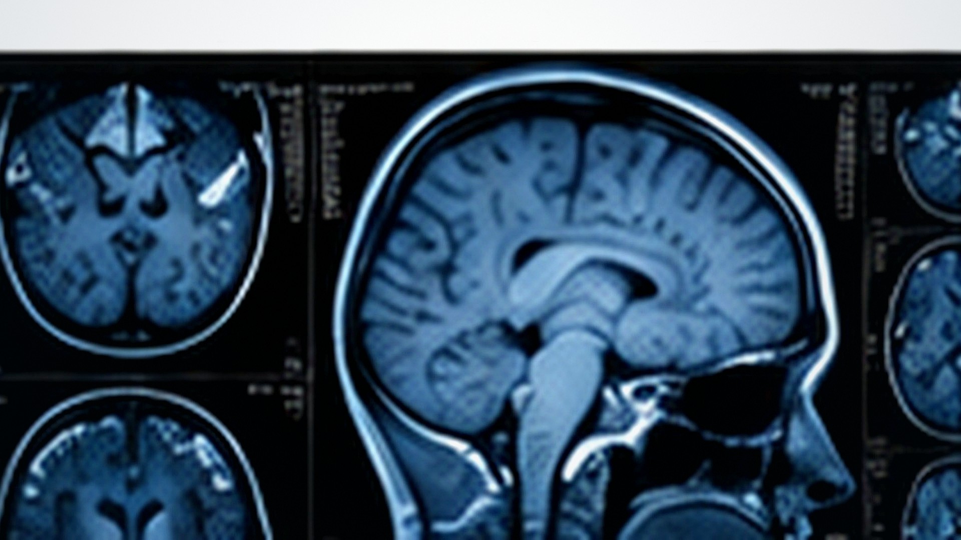
No single method can reveal every level of brain function. Electroencephalography records electrical activity from the scalp with millisecond timing. Structural MRI shows anatomy, while functional MRI (fMRI) infers activity-related changes from blood-oxygenation signals. Electrophysiology, microscopy, stimulation methods and molecular tools provide finer detail in experimental systems. Large research programs combine these approaches to link cells, circuits and behavior rather than treating one measurement as a complete picture of the brain.[4]
Blood flow is part of brain function
Neural tissue has little capacity to store fuel and depends on continuous delivery of oxygen and glucose. Cerebral blood flow is therefore tightly regulated, and sudden interruption can injure neurons within minutes. In stroke, a blocked or ruptured blood vessel disrupts that supply and can produce abrupt loss of movement, speech, vision or other functions depending on the affected network. This dependence on circulation is also why many brain-imaging methods can infer activity from changes in local blood flow or oxygenation.[5]
Functional lateralization is real but often oversimplified
The two cerebral hemispheres are anatomically similar but not perfectly interchangeable. Some language functions are more strongly lateralized to the left hemisphere in many people, while other tasks show different asymmetries. Popular claims about a logical “left brain” and creative “right brain,” however, exaggerate this specialization. Ordinary cognition depends on communication across the corpus callosum and on networks that include both hemispheres.